Revolutionizing Disease Diagnosis with Minimally invasive Optical Imaging!
Traditional tissue diagnosis requires invasive biopsies and time-consuming lab work. The Tearney Lab at Massachusetts General Hospital is changing that—developing cutting-edge, noninvasive optical imaging tools that visualize human tissue at the microscopic level in vivo. Led by Guillermo (Gary) Tearney, MD, PhD, this multidisciplinary team pioneers light-based technologies that reveal cellular structure, molecular makeup, and tissue mechanics in real time, transforming how we detect and understand disease—no scalpel required.
Slide 3
Spectrally Encoded Confocal Microscopy (SECM)
The Tearney laboratory has developed a new form of confocal microscopy, termed spectrally encoded confocal microscopy (SECM), that does not require integrated high-speed mechanical components, yet is capable of obtaining cellular-level resolution images at thousands of frames per second through an endoscope.
The Tearney Lab’s Swallowable Capsule is named one of MIT Technology Review’s Top 10 Breakthrough Technologies for 2019
We are thrilled that this year’s guest curator, Bill Gates, chose our capsule to be included in MIT Technology Review’s Top 10 Breakthrough Technologies!
“The little probe will help researchers answer questions about EED’s (Environmental enteric dysfunction) development—such as which cells it affects and whether bacteria are involved—and evaluate interventions and potential treatments”. —Courtney Humphries
Dr. Tearney’s journey as a Mass General Research Scholar is featured in the Summer 2018 issue of Mass General Magazine
“The MGH Research Scholars program offers greater freedom,” says Dr. Tearney, who was the Mike and Sue Hazard Family MGH Research Scholar from 2012 to 2017. “You can have a crazy idea that nobody believes will work, and you can try it.”
At the Tearney Laboratory, we’re proud to showcase our groundbreaking innovations in noninvasive imaging. Our portfolio highlights cutting-edge technologies designed to transform how we diagnose diseases. Each invention represents our commitment to improving patient care while minimizing invasiveness. Explore our advancements that are paving the way for a brighter future in medical diagnostics!
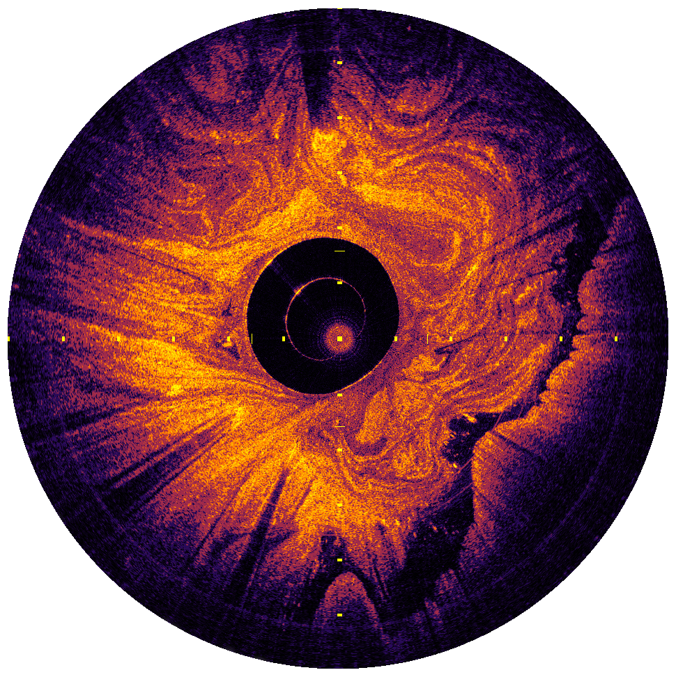
The stomach imaged with OCT Capsule
Coronary artery imaged with OCT-NIRF Catheter
Dysplastic BE imaged with SECM
Get in Touch
We’re excited to connect with you! Whether you have questions or want to learn more about our innovative technologies, reach out today. Let’s work together to improve patient care!