Dynamic OCT
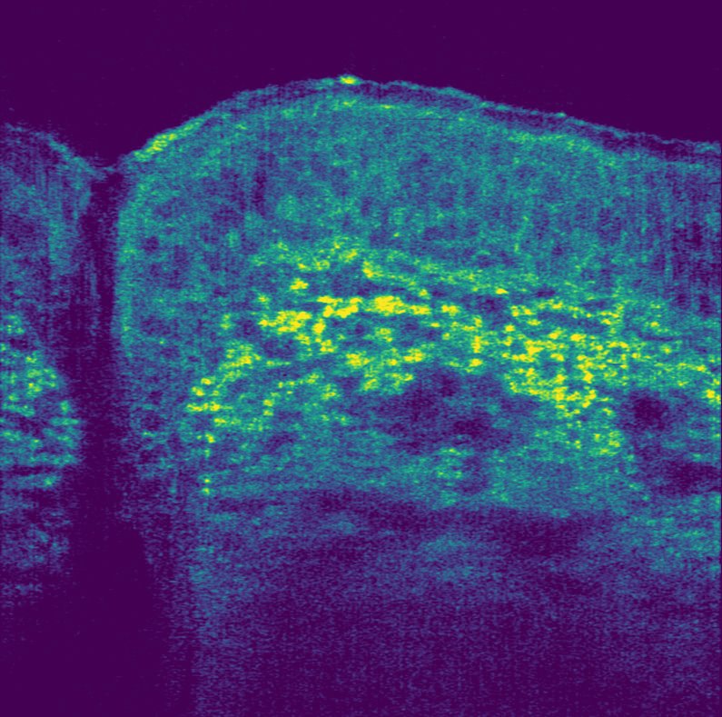
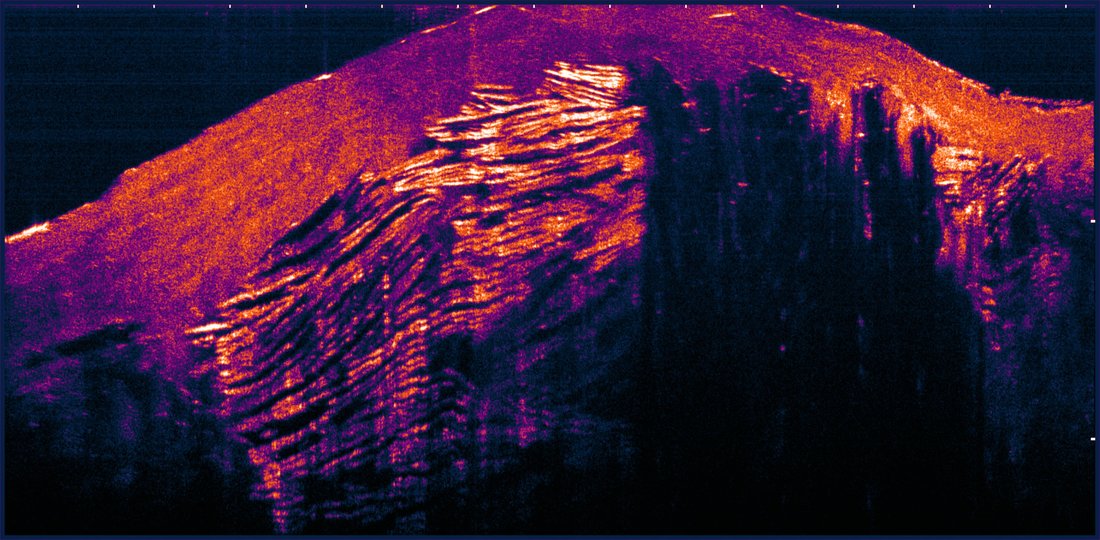
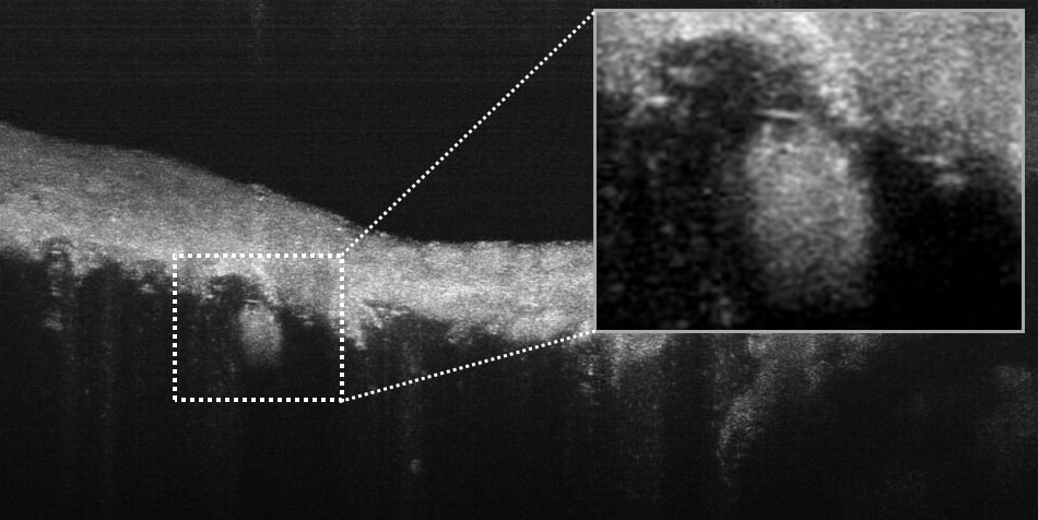
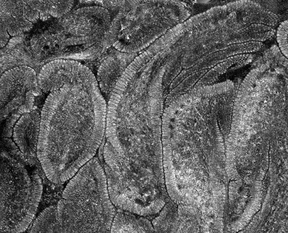
Micro-optical coherence Tomography (μOCT)
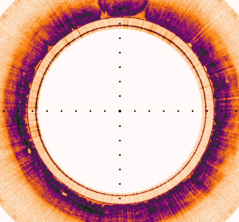
Micro-optical coherence Tomography (μOCT) is the highest-resolution cross-sectional OCT technology available today. The high resolution and imaging speed of μOCT makes it uniquely suited for imaging many biological phenomena. One such example is the respiratory airways that are lined with microscopic hairs called cilia that continuously work to sweep away mucus. This process is a crucial element of respiratory defense that becomes impaired in a variety of diseases, including Cystic Fibrosis. Another example is vascular disease where μOCT enables visualization of individual cells and their interactions, helping to discover the factors that contribute to coronary plaque formation, progression, and disruption.
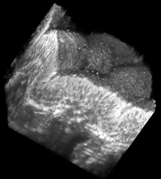

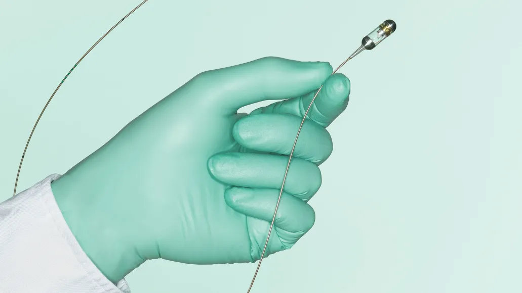
Capsule Technology
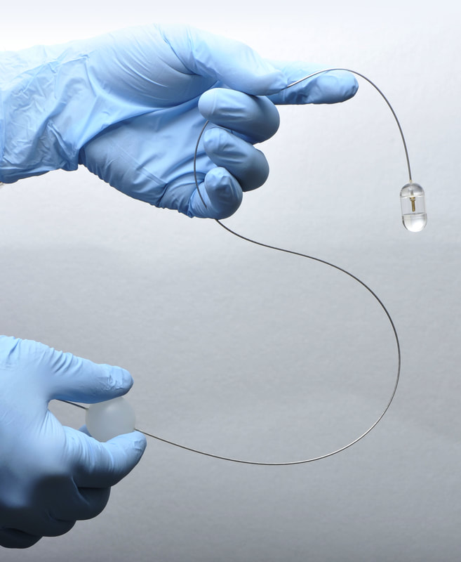
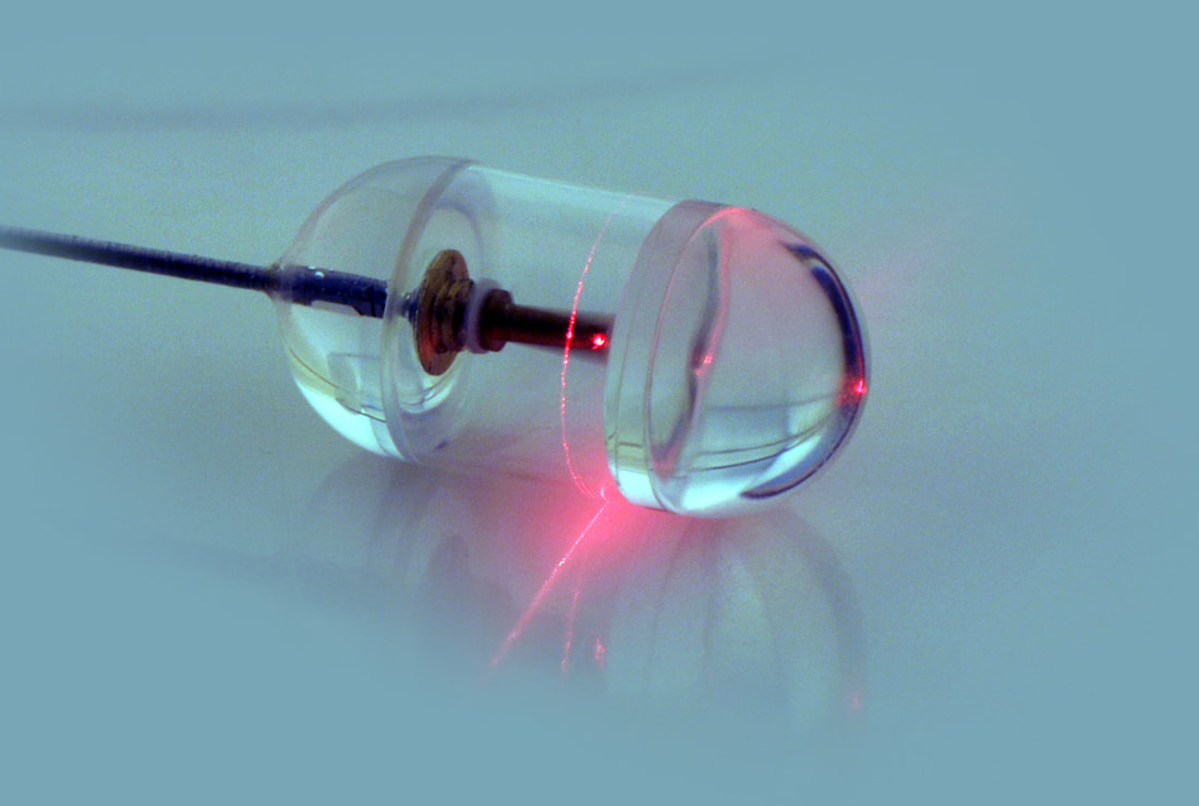
The Tearney Lab has unlocked a new paradigm for diagnosing gastrointestinal (GI) diseases that eliminates the sampling errors associated with endoscopic biopsy – all in a simple, rapid, and inexpensive procedure. The technique, termed tethered capsule endomicroscopy (TCE), involves swallowing an optomechanically-engineered pill that obtains microscopic images of the GI tract. The results obtained from unsedated human subjects showed that three-dimensional microscopic images of the entire esophagus can be obtained in just a few minutes. After imaging, the capsule is withdrawn using the tether, disinfected, and reused. The capsule is now being tested in the primary care setting to see if it can be utilized for Barrett’s Esophagus screening. Other applications being investigated include the diagnosis of Celiac Disease, Eosinophilic Esophagitis, and Environmental Enteropathy.
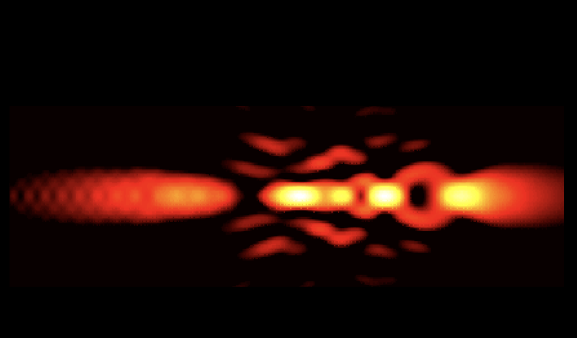
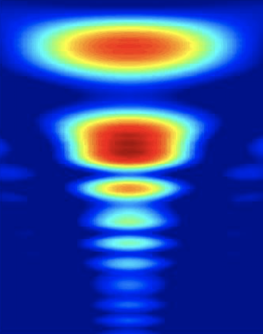
Spectrally Encoded Confocal Microscopy (SECM)
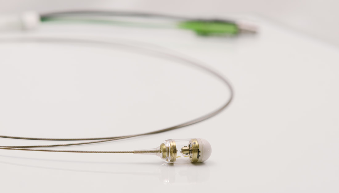
The Tearney laboratory has developed a new form of confocal microscopy, termed spectrally encoded confocal microscopy (SECM), that does not require integrated high-speed mechanical components, yet is capable of obtaining cellular-level resolution images at thousands of frames per second through an endoscope. The SECM team is fabricating endoscopic probes and capsules capable of imaging entire luminal organs with this technology.
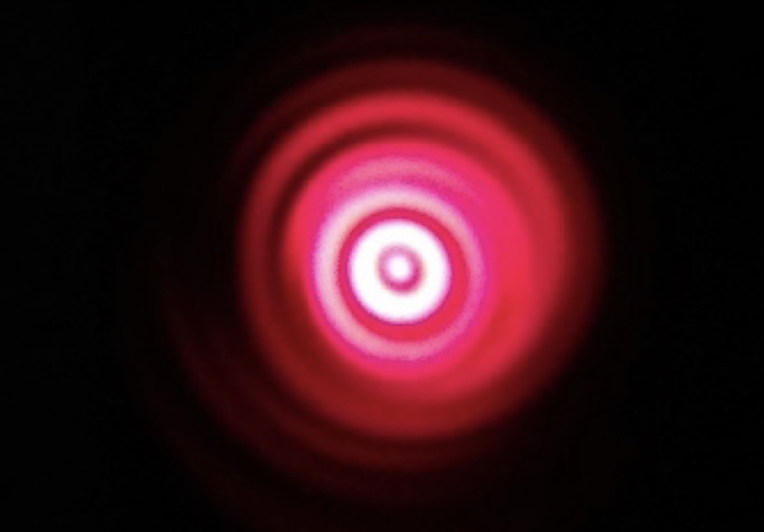
Optical Frequency-Domain Imaging (OFDI)
An advanced form of OCT called optical frequency-domain imaging (OFDI), also known as swept-source OCT (SS-OCT), achieves more than 50-fold improvement in image acquisition speed, compared to its preceding technology, time-domain OCT (TD-OCT). Our work with OFDI is focused on solving clinical dilemmas surrounding early detection of atherosclerosis and cancer.