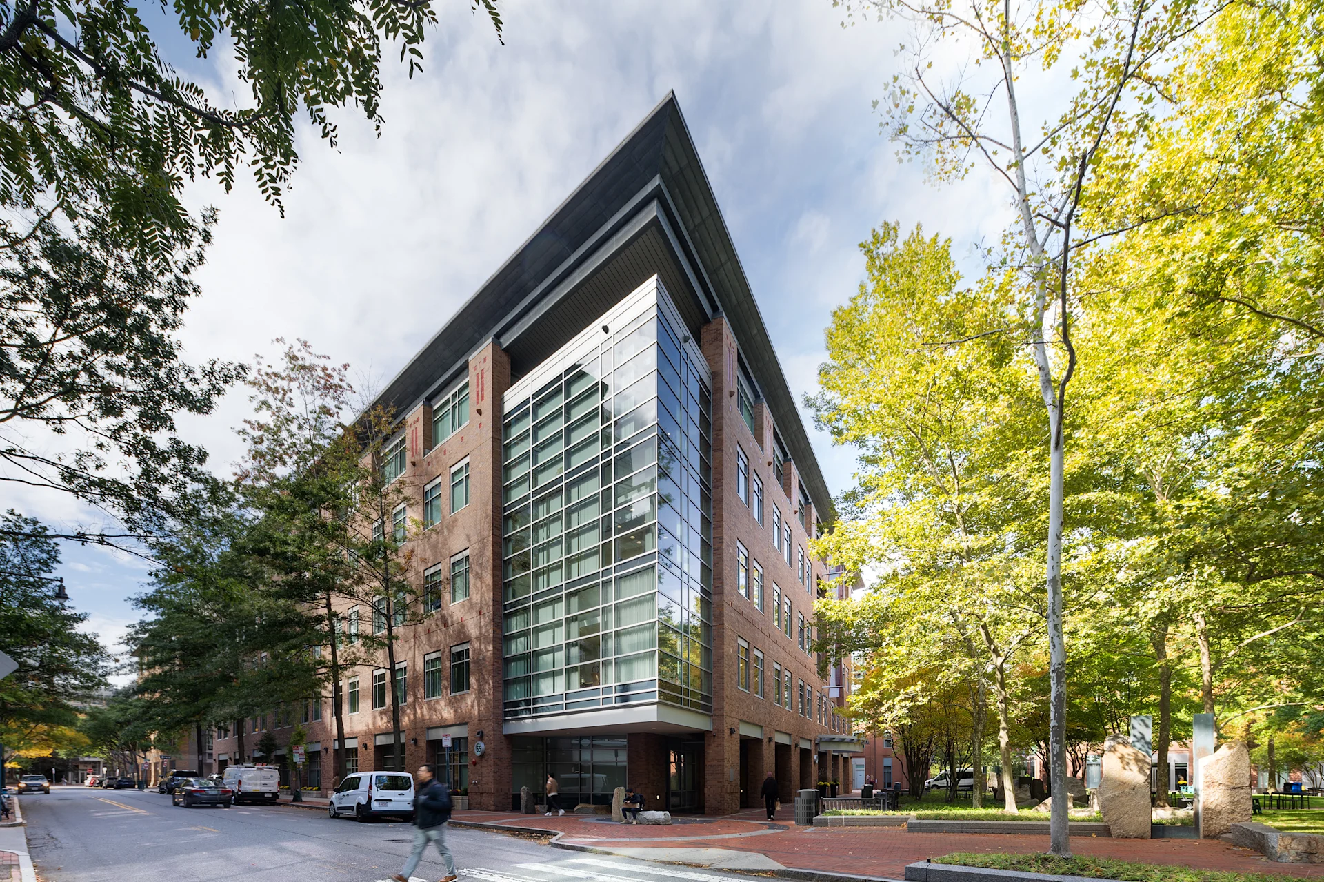
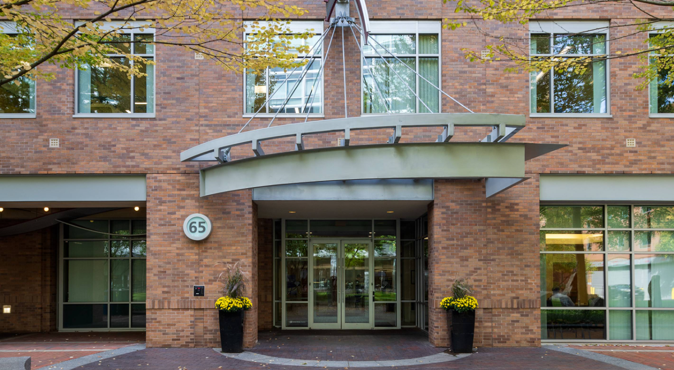
65 Landsdowne Device Fabrication Facility
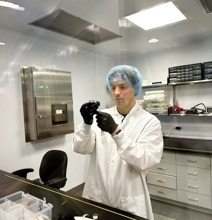
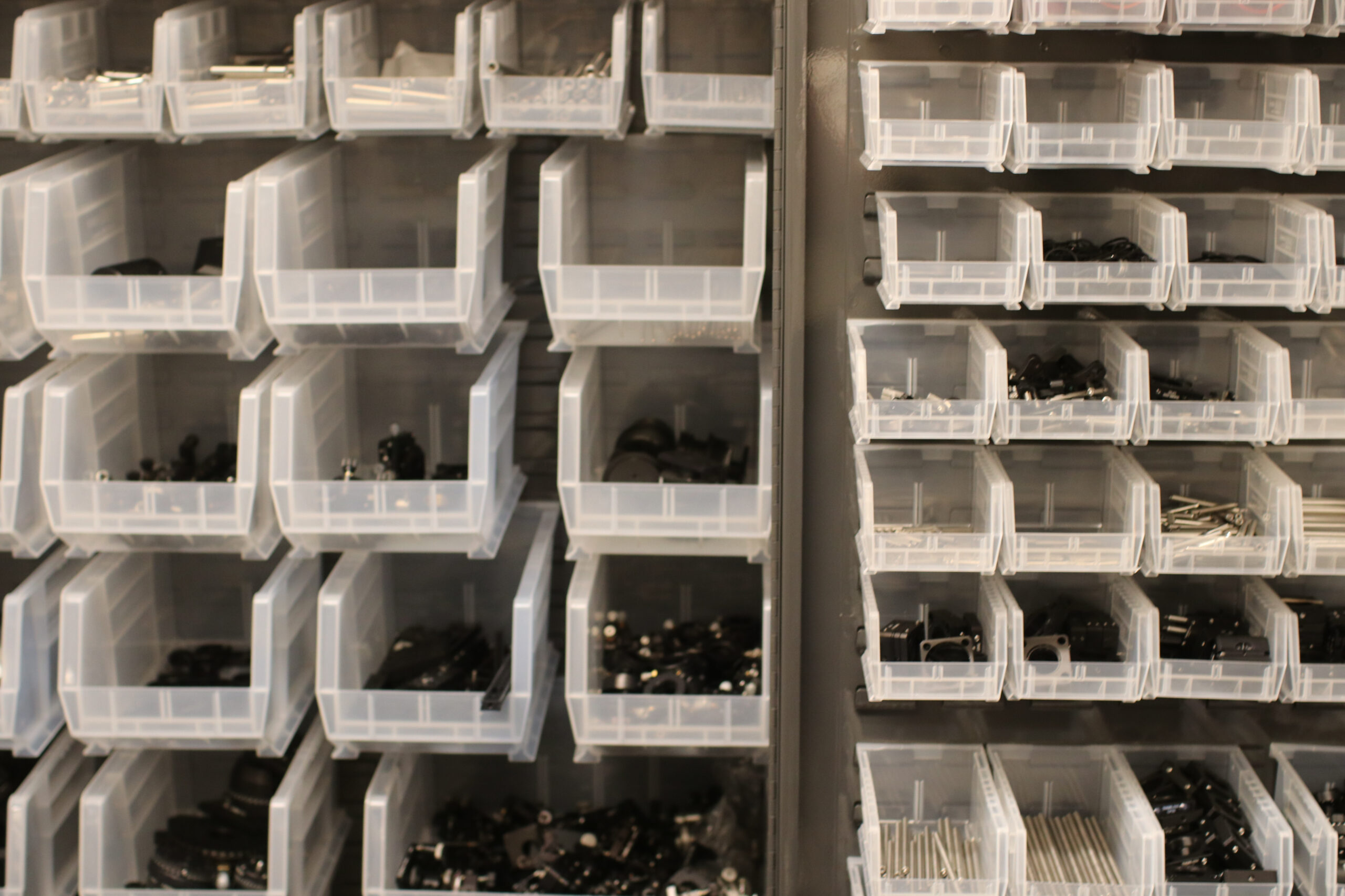
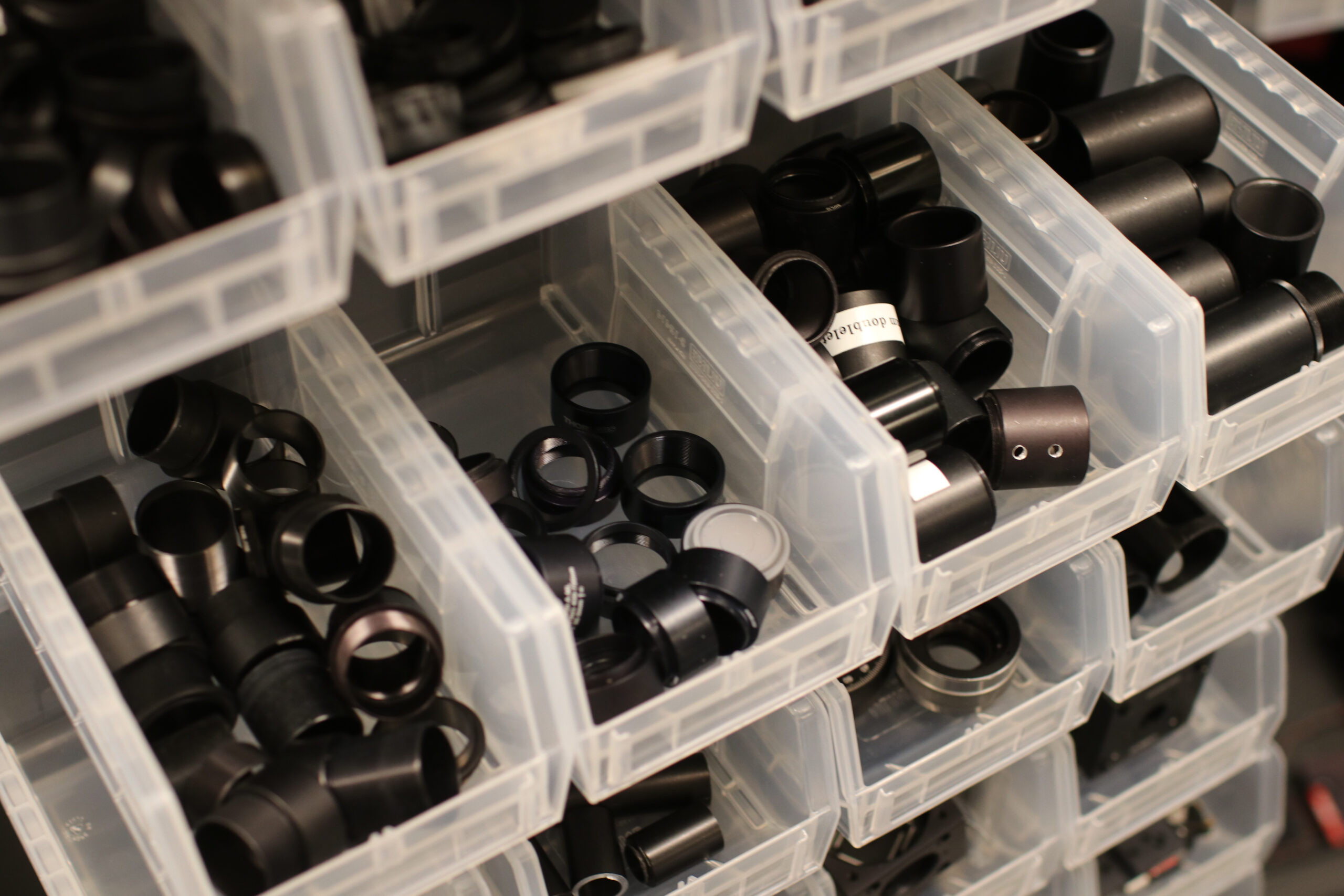
The Device Fabrication Facility comprises 3000 sq. ft. of space dedicated to GMP manufacturing of clinical devices, including gastrointestinal imaging probes and intracoronary catheters. The facility includes an optical fabrication area, 3D optics printing clean room, a state-of-the-art machine shop, two class 10000 clean rooms, and incoming inventory and finished clinical storage rooms. The Device Fabrication Facility also has seating for approximately 30 engineers and technicians that comprise the Tearney Lab’s manufacturing team.
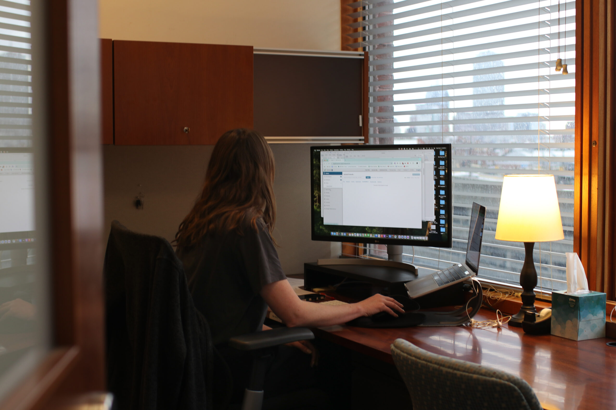
Computers. All instruments are equipped with computers for data acquisition and processing. Software programs for data acquisition (LabView, MS Visual Studio), analysis (Matlab), optical prototyping (Zemax), data visualization (ImageJ & Osirix) and mechanical prototyping (SolidWorks) are available to researchers through volume license agreements. Electronic access to journals is available through the MGH library. The laboratory maintains two secure servers for data storage. Researchers with Harvard Medical School appointments have access to the Harvard University Library System, which includes online journals and databases.
Optical Diagnostics Laboratory
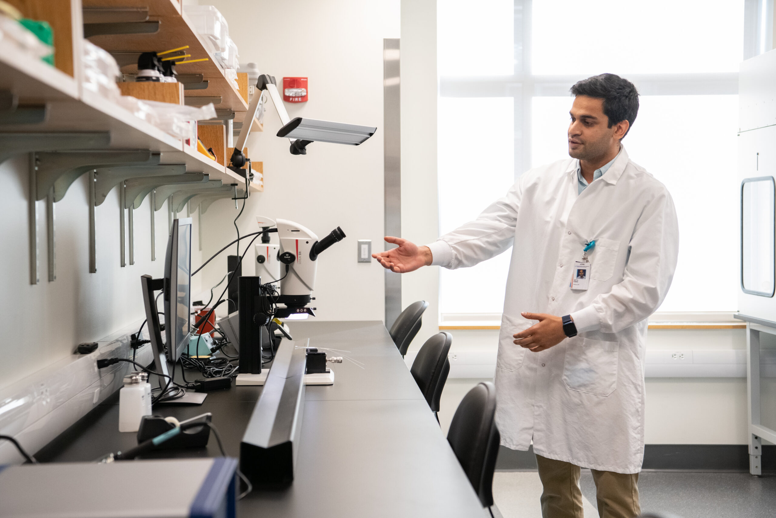
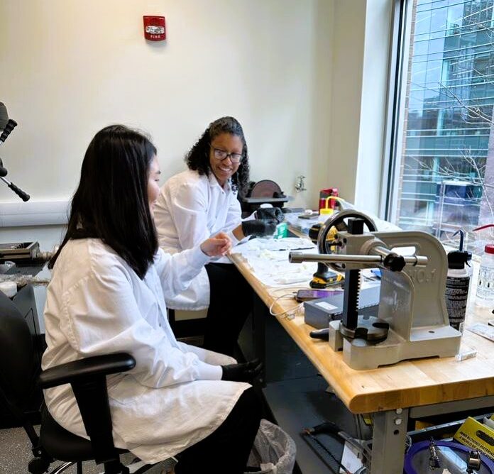
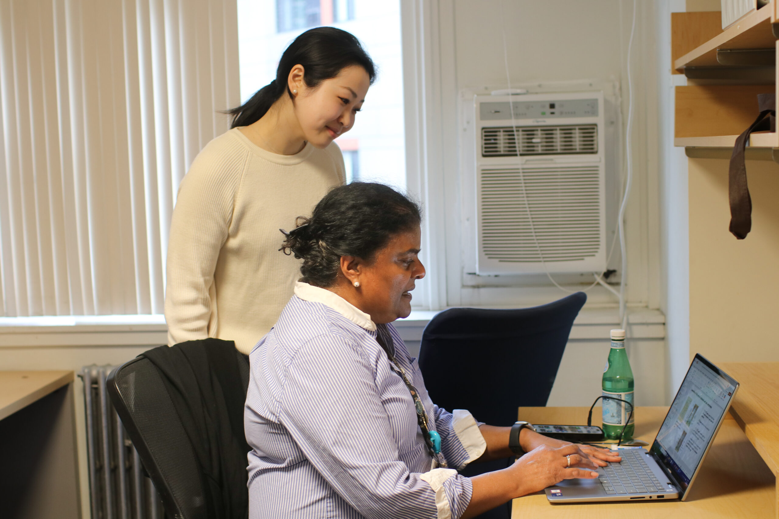
The Tearney Laboratory is dedicated to the development and clinical translation of novel optical imaging technologies. The Tearney Lab is comprised of three optics and biomedical engineering research laboratories, wet laboratory space, a dedicated pathology laboratory, cell culture room, three device fabrication facilities, a machining facility for prototyping, and a data server closet. The three optics labs are equipped with optical tables, laboratory benches, optical and optomechanical components, and electronics and optical testing equipment. Researchers share common laboratory space, facilities and major equipment.